Have you ever thought about the revolution you can be a part of when you start scanning every patient every time? Well, I did. I’ve worked with dental scan data a lot, and I’ve come to realize what we can set in motion. Imagine you can track even the slightest change in the oral cavity over the course of several years. Imagine you’d have a tool -included with your intraoral scanner- that helps you educate the patient on what has happened and what will happen if they don’t take action?
That tool exists and I am sharing my 10 commandments for how to get the most out of intraoral scanning with TRIOS Patient Monitoring - in this blog post.
1: Scan each patient upon intake to obtain a full arch baseline scan
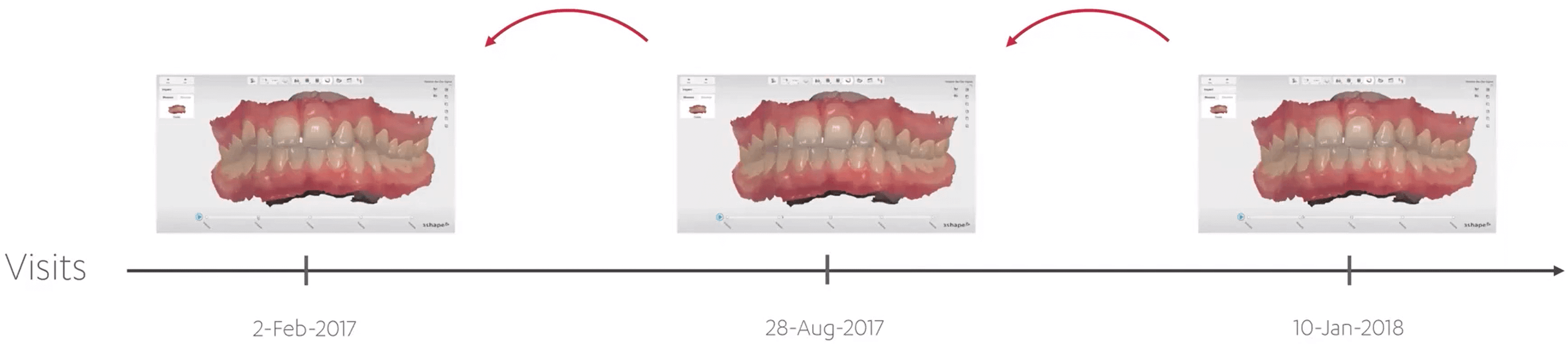
When you take a scan of every patient every time, you can truly start following the patient. However, the first baseline scan is the most critical as this will represent the reference point for your patient going forward. So it is a worthwhile investment in the future of your patients’ care.
Imagine how you could identify issues before they become problematic and intervene! Scanning at every visit lets you compare multiple scans (without limit!) over time to see how the patient’s oral health, or treatment, is developing, and you can show them their metamorphosis beautifully on screen. And the best part is: taking a scan and ensuring it gets filed properly takes less than 5 minutes.
2: Ensure to clean and dry the patient’s mouth before scanning
Scanning a patient directly in the mouth has its own principles and pitfalls. It’s not rocket science, and we’ve outlined it in our scan strategy onboarding (requires 3Shape Account) and video tutorials and webinars. One of the key things I would recommend in order to get clear and accurate scans is to always clean and dry the mouth before scanning. Saliva, or leftovers from lunch, can for example impact the shape, color and reflection of the scan making the scan less valuable for comparison and monitoring down the road.3: Ensure that scans contain a minimum number of holes and sufficient color data
When scanning, it is important to ensure you capture as much data as possible- quantity and quality are both important. After all, you don’t know where in the patient’s mouth future changes or disease markers will emerge. Keep in mind that any holes or insufficient color data will impact the quality and reliability of the scan.
The TRIOS scanning software highlights holes in the scan as part of the scanning process- make sure to rescan those sites. Also, the blue-overlay tool used with shade measurement will give you an indication of which areas contain insufficient color data.
4: Scan using the 3Shape scan strategy for full arch scans
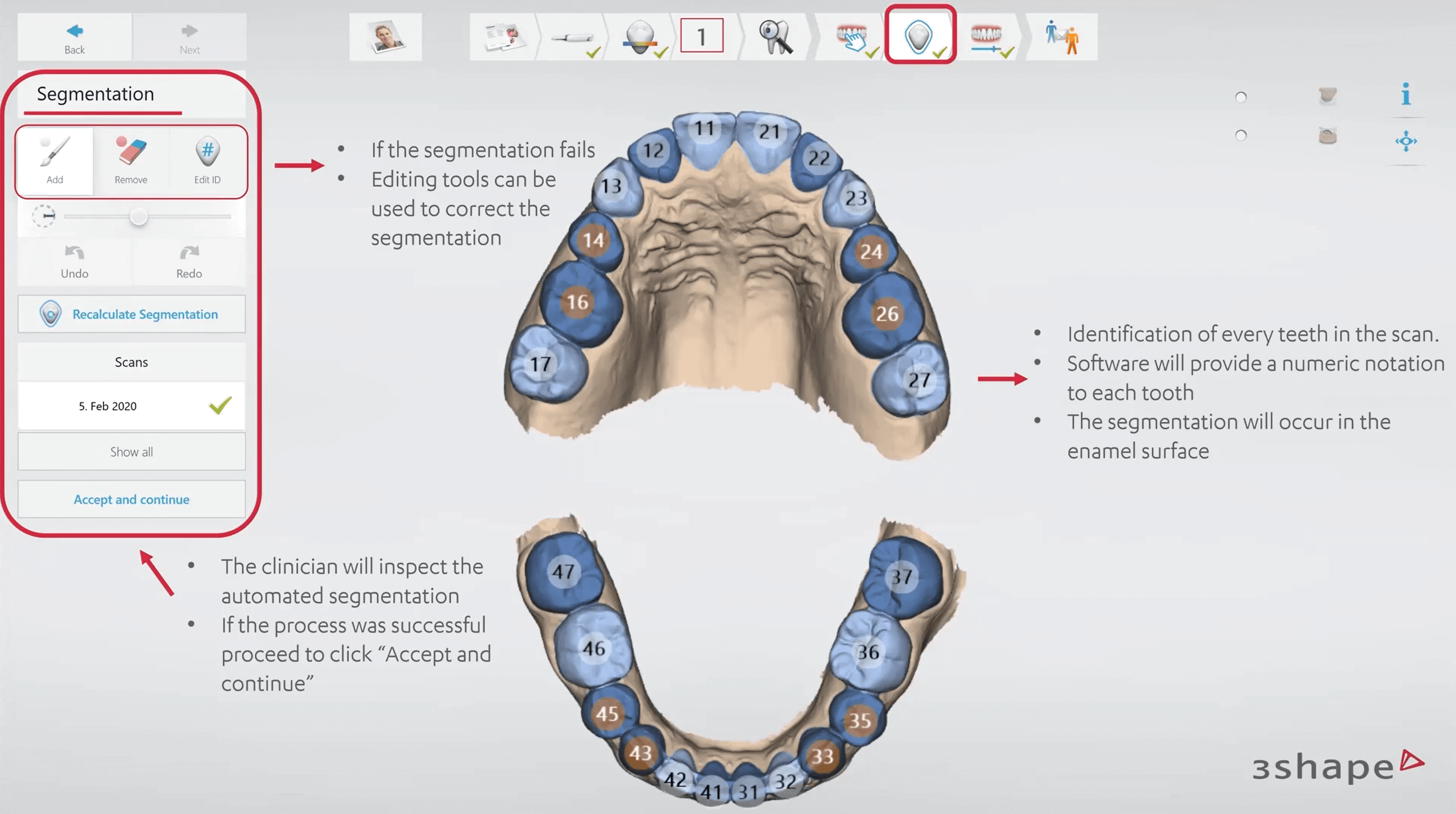
When you follow our designated scan strategy for a full arch scan, you can be sure to have captured everything including the bite. The recommended scan path consists of 3 sweeps: occlusal, lingual, and buccal to ensure good data coverage of all surfaces. The best scanning method is to start with the molar occlusal surface. This has greater details for easier identification. Everything else will be done by the software: segmentation, identification of every crown or lacking teeth.
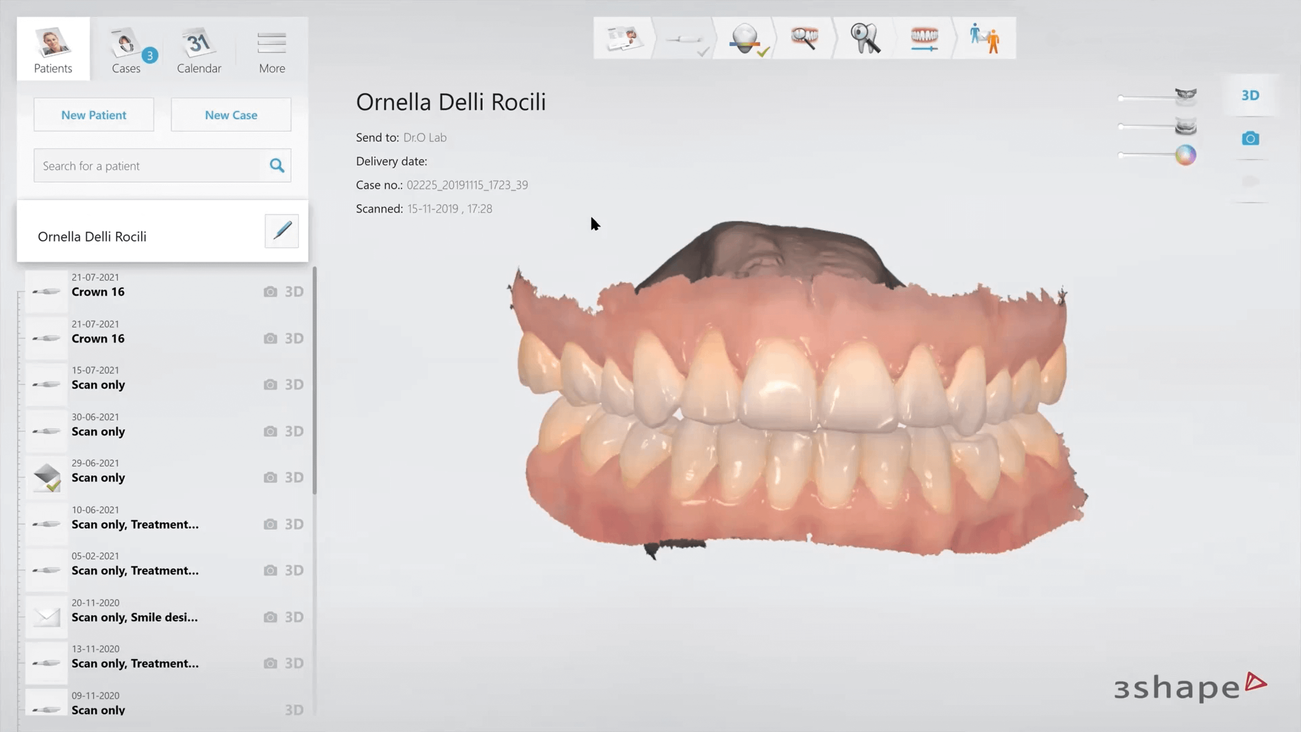
5: Scan using the same digital patient for each follow up scan, rather than creating a new patient each time
From a data point of view, I recommend you ensure you create only one patient and store all their scans under that unique patient ID. Sometimes, patients get entered twice, or more, by accident, or a typo occurs and then the patient’s precious scan data gets distributed over multiple entries. When this happens, we need to clean and tidy up - and who has time for that?! This might sound like a no-brainer, but I have seen practitioners do this differently many times. Especially in practices where multiple people operate the systems, this can go wrong.
6: Re-scan patients on a yearly or biyearly basis, or as is otherwise appropriate
Taking scans regularly means you keep a record of how the patient’s oral cavity has evolved over time. I recommend capturing data every year or even twice a year, depending on your patient and their needs and risk factors. The way to ensure this doesn’t eat up all your data storage capacity, is by trimming the scans so it doesn’t consume as much memory, and ensuring you compress your scans.
7: Backup your patients’ scans regularly, both onsite and offsite
Anyone with a computer will acknowledge: it is not a nice experience to have data stored only on one computer and then have that computer crash. Even though we trust our systems, you never know what happens to a computer, a hard drive, or any other data storage device you might use. That’s why we recommend you work with a backup routine, every day, week or month. Scans really don’t take that much memory and can even be compressed (they don’t take more than 3 to 10 MB which is the size of an average photo).8: Check and verify the segmentation of the scans
Segmentation is always the first step after scanning. During segmentation, the teeth are separated from the gingiva and labelled based on a chosen tooth numbering system. The software does this automatically, but each scan needs to be carefully inspected and corrected. Automatic segmentation is currently optimized for scans of unaltered adult teeth – for any other teeth, you can perform segmentation manually. Missing teeth will be highlighted by the adjacent teeth IDs.9: Endeavour to understand patient monitoring as a tool, and interpret its results with an awareness of its strengths and limitations
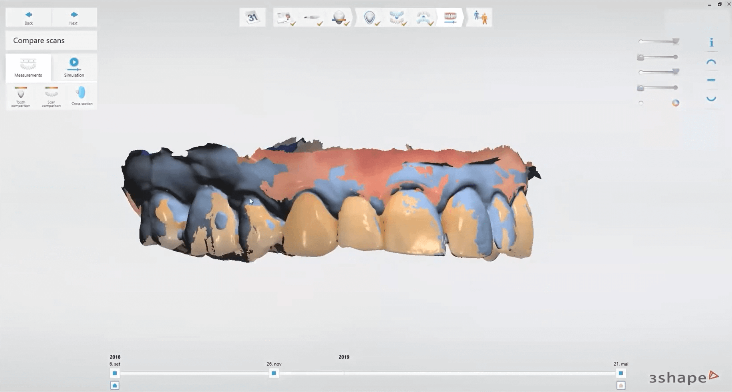
Software to help you monitor your patient is no more than a tool: it lets you capture, visualize, and compare data. There’s only so much the machine can do to interpret the data, so your role as a dental practitioner is to utilize your years of education and experience to guide and advise the patient in their dental health journey. You can make the difference by explaining what has happened, what is at risk of happening, and how the patient can turn the tide.10: Use patient monitoring to the benefit of your patients – to educate, engage and prevent
A digital representation of how your patient’s oral health evolved over time is an excellent way to show your patient how severe (or not severe) their case is. Its visualization lets you explain how treatment can help them and showcase how their progress goes. That is why my last commandment puts the patient center stage: it is ultimately about their wellbeing and health.
Want to get the full walkthrough of how to scan, what the benefits are, and how to get the most out of TRIOS Patient Monitoring? Our in-house doctor Ornella gives you a complete walkthrough in this 1 hour webinar.