A lot is happening in the world of intraoral scanning. In my four years as a Global Training & Application Specialist, I have spoken with an endless number of dentists that were just getting started with their digital journey. In that regard, I’ve seen a few characteristics make or break dental workflows.
Here’s the 7 intraoral scanner features that I think you should not compromise on when you start scanning your patients.
#1: Versatile enough to help you through a busy day
Let’s do a mind exercise: let's say you got yourself an intraoral scanner. It’s wireless because you like the flexibility it gives you. You are maybe not scanning every patient every time yet, but you are close to. And then, suddenly, you have a big day in the clinic with many, many patients. They just keep coming. And you keep scanning.
Then, you realize you forgot to charge the batteries of your scanner. By accident. What happens? You don’t want to be without a scanner. That’s why I recommend you look out for a solution that allows for versatility, both in terms of cords and wireless options, but also in terms of how easy it is to take the scanner from one room to the next. And in terms of how easily you can show, play, visualize and manipulate in 3D on the screen. In front of the patient, so they can see your visuals. Preferably in a way that is ergonomic for you so you can work with them either standing up or sitting down.
Then, the tip. The tip needs heating in order for it to be ready to scan. So that takes time. And also battery life. If you can get your hands on a scanner that heats up in just a few seconds so you’re scan ready while preparing the patient, then that can also save you a lot of time.
#2: Artificial Intelligence as an extra pair of hands
Something else that is fairly new in the world of digital dentistry is artificial intelligence (AI). AI has proven to make dentistry easier, but it is important to ask yourself whether the scanner you have in mind utilizes artificial intelligence? And if it does, how does it actually work?
I know a lot of dentists don’t know yet how far AI can go and how it can help in the dental world, so let me explain in simple words what it can do. Labs can for example automate their crown designs in a matter of seconds. Or move soft tissue such as lip, tongue, cheeks so you have a perfect starting point to plan your treatment.
The arrival of AI makes it possible to let the scanner do all the manual work for you.
On one side of the screen you can see how it looks without AI. The other half shows you how the scan is cleaned up nicely with AI technology. It makes it easier for you to do a very clean, very nice and fast scan.
#3: Easy on children (and adult patients)
As a doctor, you work for your patients. You want to ensure they are comfortable. There are many adults who suffer from dental anxiety and even more so for children, dental visits can be a challenge. How many times have you had a crying child in your practice when they had to get their impression taken? Nobody likes to have an impression made, no matter how expensive the material is, the flavor or scent you use. You can use aromatherapy in the room to make the experience more ’comfortable’ – but it will never become truly pleasant.
It’s not hard to imagine how excited kids will be when they can just see their own mouth on a screen. A funny story actually comes from Dr. Ramya Kannan from 3Shape Academy India. Here is a scan that she performed on her nephew, a three year old toddler.
After the scan, the child said that ‘auntie Ramya took a selfie of my teeth’.
Think of how an intraoral scanner can turn the experience around for children like that. For me, these stories are priceless.
But then, of course, let’s not forget about our beloved adult patient demographic. How can your scanner turn a dentist visit into a pleasant experience for that audience as well? Tools to monitor the patient’s mouth over time, for example, or to visualize their treatment in advance, can really help make your treatments into a pleasant experience.
Because do you remember exactly what the tooth looked like three or six months ago when the patient was there? In these situations, it is valuable to have the ability to superimpose multiple scans and then show the outcome or the difference between the surface enamel. It allows you to quickly see small details that are not visible when looking at them with your eyes. As a clinician, you can then evaluate chemical or mechanical erosion of enamel and measure it by the millimeter!
Another thing to be on the lookout for with a scanner is the option to simulate patient’s potential smiles. Vivid and realistic renderings of reality, superimposed to the patient smile and just showing them how great they can look are a fantastic acceptance booster. It means a lot for patients to be able to see themselves.
#4: Dynamic bite registration
Motion is yet another of these things that you need to be aware of when getting a scanner. Not all scanners are actually capable of doing dynamic bite registration. What you want is that after you’ve scanned the upper and lower jaw, static occlusion, you can add mandibular excursions. So, you put the scanner in the patients mouth in static occlusion, turn on the scanner and record the protrusion, retrusion and laterotrusion.
Then, you can evaluate and perform diagnostics based on these contact points. Also, the ability to share this with your lab allows your dental technicians to use this info to design, adapt your crown, or any other restoration in your treatment plan. This will ensure a perfect fit of your crown, with minimum to none occlusal adjustment since the lab used the patient’s motion to adapt the design. Cool, isn't it?
#5: Color scanning
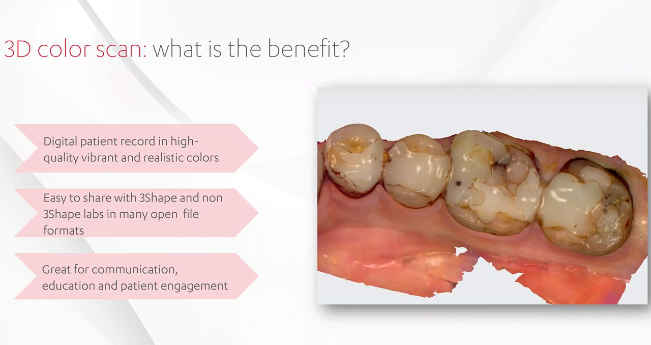
Another thing is color. Back in the days monochrome was still the standard, but intraoral scanners have gotten better and better in their color representation. As a GP or as a dental technician you can really benefit from this. Of course, when you have the patient on the chair, color is a great communication tool because you can perform a diagnostic scan and show their oral cavity in 3D and real colors. These scans will also help in the conversation between clinician and patient. Think about it: you will have current oral health status, oral hygiene, preparation assessment and selection of enamel and dentin shade, all in one place!
However, let's not forget how the labs also benefit from this. Dental laboratories rely on the shade selection of the dentist. In this case both lab and doctor will have the same information, so communication will definitely be improved! It ensures predictable outcomes for the doctor and perfect aesthetics for the technician.
Let's take an example: when I'm performing a crown, I want to make sure that I know the exact shade details of my patient. It requires a shade measurement tool that allows me to detect the shade not only from the enamel, but also from the dentin. And I want that tool to support the industry’s most used shape measurement scales from from Ivoclar Vivadent to VITA. Not only do I want to be able to visualize it myself - I also want to quickly be able to send it to my lab. So that they will be able to perfect aesthetics with the same shade that I read on the clinical side.

When you look at this rendering of color in the scan, you can see how it helps: you can see all the different micro leakages on those restorations. See how this will be an amazing diagnostic aid tool? To visualize and talk to the patients and say, what is the treatment plan?
#6: A cloud solution that brings everything together
When working with analog models, there is a risk of contamination and infection. And there's also the risk that the model's a bit more brittle and can break accidentally. You don't have that when you store your 3D models in the cloud. Ideally, you work from one platform that serves as your desktop and gives you easy access to all the different steps in your workflow. There's no risk of contamination and -when you use a properly built cloud solution- you can bring in scans that you have taken long ago. And you can export them into open file formats, which is the industry standard - or use a file format that includes the colors as well. And then you can send, of course, to other colleagues, or straight to the dental laboratory. You want the platform for your digital dentistry to be easy to use. Any new user should be able to go into the software and start scanning.
#7: Connectivity with your dentistry ecosystem
The final thing that you should consider as vital to your digital dentistry solution is to what extent it is open. In essence, there are two ways of approaching your digital dentistry investment (just like with investing in a computer, for example): do you want an open or closed system? And then what about the ecosystem that you can work with? How does the scanner interact with the different providers? And how does the ecosystem support your own daily routines?
A good reason to choose an open ecosystem is the connectivity with labs or other partners around the world to send your cases to. Some of them will of course only offer specific things, but others will be offering a wide range of different treatment options including sleep appliances, clear aligners or implants. An ecosystem like that means that you always have the option to grow from your regular day-to-day work and expand to bridgework and then implants, and so on. It allows you to transition from what you've been doing, grow from analog to digital, and then you can expand your possibilities because the tools are always at your disposal.