- Home
- Blog
- Explore treatments
- How does digital dental monito...
How does digital dental monitoring work?
Upon sitting in the chair, the dentist asks, “How does your mouth feel? Does anything feel different?” The patient reports, “Everything feels okay, I think, but sometimes I feel like my teeth are shifting. And by the way, are my gums receding up here?” Sounds familiar? In fact, we hear things like this from patients all the time. But how can we tell if something has changed?
How to identify small oral health changes at an early stage?
For the majority of my 16 years in practice, when patients come in for their bi-annual recall visits, we dentists have relied on radiographs (x-rays), visual examination, chart notes, and photographs to follow and manage a patient’s oral health. And the data acquired with these methods is still a very important component of our records. However, when small changes occur in the mouth over the years it can be difficult to definitively quantify, measure, compare and identify them at an early stage.
Although annual radiographs can be compared from one year to the next to assess cavities and jawbone levels, there are many other variables that are not visible on a two-dimensional x-ray. Including, but not limited to, soft tissue changes, tooth shifting, tooth wear and gingival levels (gum tissue positions).
Getting data without radiation or fear of gagging
An emerging paradigm shift occurring today is the use of digital intraoral scans to create objective data sets annually that can be analyzed by the dental team to measure changes in the mouth. These scans are obtained without the use of radiation, traditional impression material or fear of gagging from the patient’s perspective.
With the introduction of powerful new dental software that can align scans taken over time, we can now make very detailed analyses and comparisons of the mouth in a way that was impossible even one year ago. The uses and educational opportunities these software programs provide for us are now being realized and discovered on practically a daily basis.
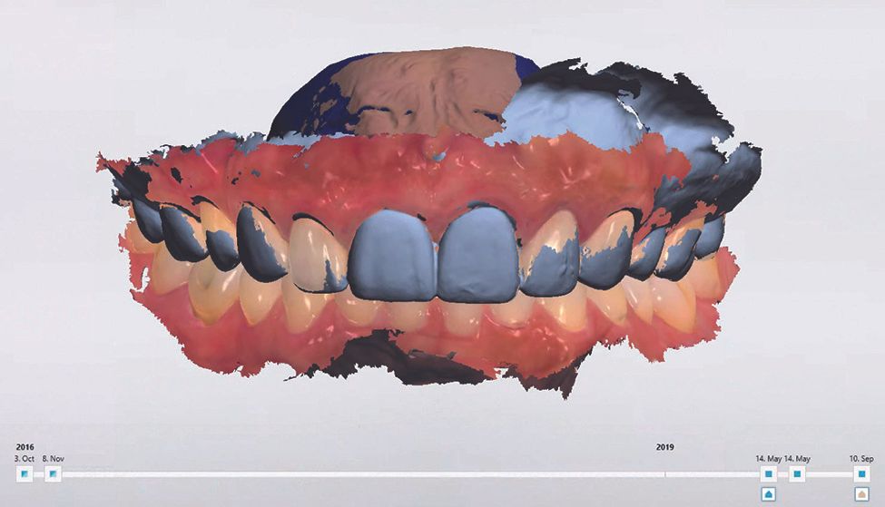
Video timeline that shows actual changes over time
Now when a patient asks, “Doctor is my bite changing?” or “Are my gums receding?” we can measure to within a tenth of a millimeter to assess changes and report findings to the patient. In addition to being able to visualize the scans stacked upon each other, we even have the ability to create a video timeline and actually watch the mouth changing over time.

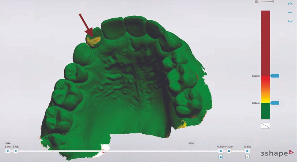
We can use the difference maps where anything colored green is stable while anything yellow and red has changed. Furthermore, we can slice the 3D scan in any direction and make 2D cross-sectional measurements and analyses.

Monitoring oral health at an entirely new level
Our lives are increasingly filled with data and digital technologies these days. We’ve come to expect to have vital signs and bloodwork taken and compared in digital medical software at our routine visits with our physicians. We can soon expect to have a digital intraoral scan in addition to radiographs at routine dental visits to obtain crucial data to detect, identify and discover subtle changes in the mouth sooner than ever before. This will allow dentists to diagnose problems earlier, treat more conservatively and monitor oral health at an entirely new level. The future of dentistry is occurring now thanks to vast improvements with integrated digital technologies.
About Dr. Naren Rajan
Naren Rajan, an honors graduate from Rutgers School of Dental Medicine, is a leader in the education of digital techniques in dentistry and lectures nationally on the topic. In practice for over 15 years, Dr. Rajan focuses on incorporating digital techniques into all aspects of general practice. He believes in finding the best combination of traditional techniques and cutting-edge advancements to improve patient outcomes. More about Dr. Naren Rajan.
Dr. Najan uses 3Shape TRIOS in his dental practice.